


What is Skin Cancer Surgery?
For skin cancers (basal cell & squamous cell), the most specialized and advanced surgery is Mohs micrographic surgery. Named after Frederic Edward Mohs, the surgeon who developed the technique, Moh’s surgery is the “gold standard” for removing skin cancers.
What is MOHS Micrographic Surgery?
New treatments for skin cancer are appearing and evolving rapidly in recent years. However, one surgical technique has more than stood the test of time. Developed by Dr. Frederick Mohs in the 1930s, Mohs micrographic surgery has, with a few refinements, come to be embraced over the past decade by an increasing number of surgeons for an ever-widening variety of skin cancers.
MOHS Micrographic Surgery is a highly specialized procedure used in the total removal of skin cancers. This surgery uses a layer by layer method of removing skin cancer, with microscopic examination of nearly 100% of the removed tissues margins. This procedure yields a cure rate of over 98%.
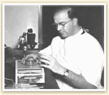
After a layer of tissue is removed by Dr. Nguyen, it is taken to our in-house laboratory. The removed tissue is precisely marked with special inks by our histo-technician who is specifically trained in the MOHS technique. A Micrographic map is made which corresponds to the excised tissue. The tissue is then frozen and slides are produced and examined by Dr. Nguyen.
If no evidence of skin cancer remains, the procedure is complete. If the area is still positive for skin cancer, the precise location is marked on the map, and the physician takes another layer on just the positive areas(s). The same process of inking, freezing, cutting, staining and reviewing of slides is repeated until a cancer free layer is reached.
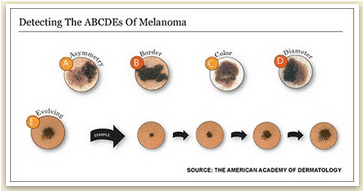
In the past, Mohs was rarely chosen for Melanoma surgery for fear that some microscopic melanoma cells might be missed and end up spreading around the body (metastasizing). However, efforts to improve the Mohs surgeon’s ability to identify melanoma cells have led to special stains that highlight these cells, making them much easier to see under the microscope. Thus, more Mohs surgeons are now using this procedure with certain melanomas. With the rates for melanoma and other skin cancers continuing to skyrocket, Mohs will play an ever more important role in the coming decades.
Today, Mohs surgery has come to be accepted as the single most effective technique for removing Basal Cell Carcinomas and Squamous Cell Carcinomas (BCCs and SCCs), the two most common skin cancers. It accomplishes the nifty trick of sparing the greatest amount of healthy tissue while also most completely expunging cancer cells; cure rates for BCC and SCC are an unparalleled 98 percent or higher with Mohs, significantly better than the rates for standard excision or any other accepted method.
The reason for the technique’s success is its simple elegance. Mohs differs from other techniques in that microscopic examination of all excised tissues occurs during rather than after the surgery, thereby eliminating the need to “estimate” how far out or deep the roots of the skin cancer go. This allows the Mohs surgeon to remove all of the cancer cells while sparing as much normal tissue as possible.
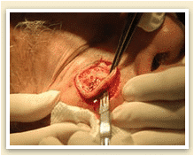
The procedure entails removing one thin layer of tissue at a time; as each layer is removed, its margins are studied under a microscope for the presence of cancer cells. If the margins are cancer-free, the surgery is ended. If not, more tissue is removed from the margin where the cancer cells were found, and the procedure is repeated until all the margins of the final tissue sample examined are clear of cancer. In this way, Mohs surgery eliminates the guesswork in skin cancer removal, producing the best therapeutic and cosmetic results.
How long does the procedure take?
It is recommended that all MOHS patients allow adequate time to complete the procedure without interruption. Depending on the size and shape of the tissue layer, each layer may take 30-60+ minutes to process. Therefore, MOHS surgery requires patience by both the patient and the physician
Learn more about Mohs Micrographic Surgery
To learn more about Mohs Micrographic Surgery or to schedule surgery, you may call our surgery scheduler at (714) 531-2966. For additional information on skin cancer and MOHS surgery, please consult some of the organizations of which we are members:
American Academy of Dermatology – https://www.aad.org
American Society for Dermatologic Surgery – http://www.asds-net.org
The American Society for MOHS Surgery – http://www.mohssurgery.org
Skin Cancer Foundation – http://www.skincancer.org
Want Beautiful, Clear Skin?
It starts here. Come see us today!